Fig. 4.

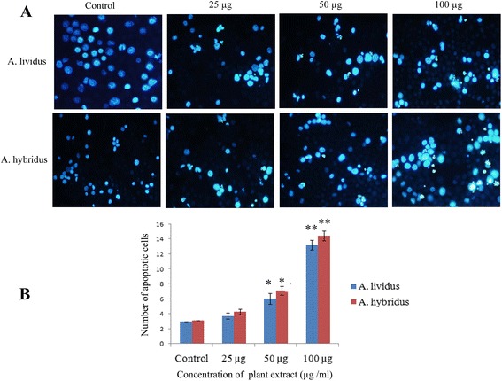
Detection of apoptotic cells using DAPI staining after six days of treatment. a Treatment was started after 24 h of EAC cells injection, at the concentration of 25, 50 and 100 μg/ml. Marked apoptotic features such as membrane blebbing, cell shrinkage, chromatin condensation, aggregation of apoptotic bodies and brightly stained nucleus under blue fluorescence etc were observed in the treated groups, in contrast to round shaped and less brightly stained control cells. b Number of apoptotic cells per side was estimated by counting apoptotic cells in five different fields. Each value represents as mean ± SD (n = 3). Significance was set at P <0.05 (*) and P <0.01 (**) with respect to control
