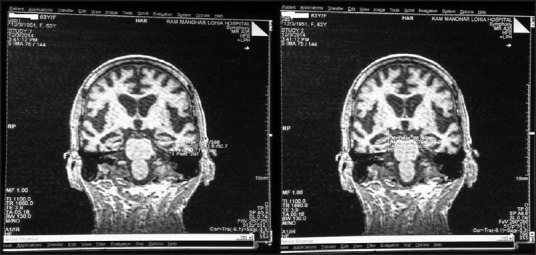
Figure 1.

MRI of brain showing oblique coronal images. ROI approach showing the area of hippocampus highlighted in T1-weighted images. Area in the consecutive slides has been summed up by manually outlining hippocampal area and multiplying by interslice gap and slice thickness to obtain the volume in cubic centimeter (cm3)
