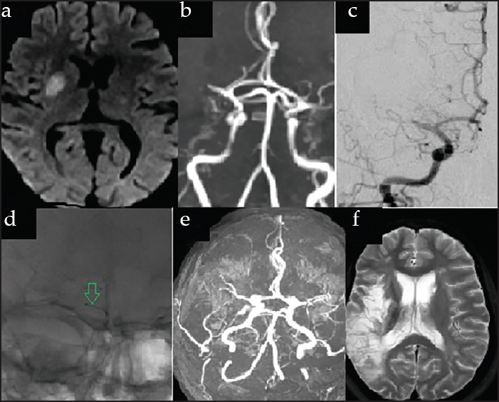
Figure 2.

(a) DWI MRI images showing a well defined infarct involving the right putamen, (b) MRA and (C) DSA showing cut off of the right MCA in the proximal M1 segment, (c) The PENUMBRA Catheter (green arrow) deployed in the occluded segment (D) follow MRA showing partial recanalisation of the right MCA (d) T2 axial image after 1 month showing extension of infarct to involve only the territory of the superior branch of the right MCA
