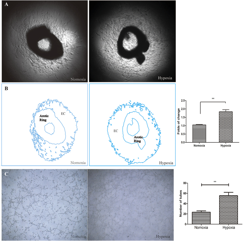
Figure 2. The effect of hypoxia on angiogenesis was assessed using mouse thoracic aortas and HUVECs.
(A) Aortic ring assays revealed increased EC proliferation under hypoxia. (B) The outline in (A) was drawn using Adobe Illustrator software, and the area was calculated. (C) Representative images of tube formation under normoxia and hypoxia. Tube formation was enhanced under hypoxia for 6 h. (EC: Endothelial Cells) **P < 0.01 vs. normoxia.

