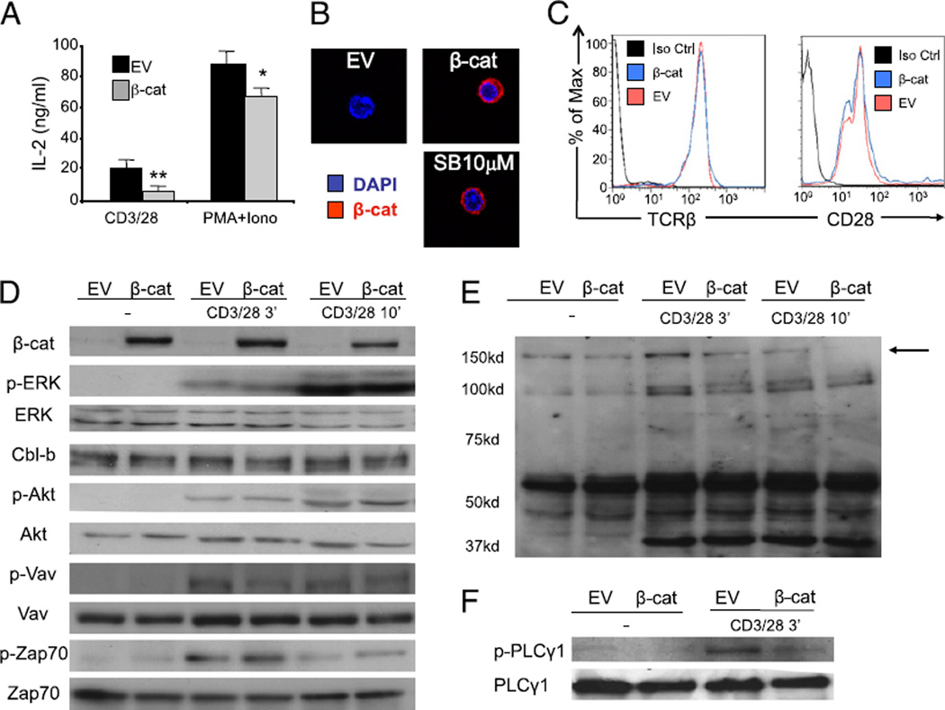
FIGURE 4.
Expression of stabilized β-catenin in T cells inhibits proximal TCR signaling. Purified peripheral T cells from CAR Tg mice were transduced with an EV or an adenoviral vector coding for a nondegradable form of the β-catenin (β-cat). A, Transduced cells were left untreated or stimulated with either anti-CD3 and CD28 or PMA and ionomycin, and IL-2 production was measured by ELISA 16 h later. An average of four independent experiments is shown. B, EV or β-cat transduced T cells were analyzed by confocal immunofluorescence microscopy to determine the localization of β-catenin. The nuclear dye DAPI is shown in blue and β-catenin in red. A representative image of highly positive cells is shown (original magnification ×400). C, Transduced cells were analyzed by flow cytometry for the expression of CD28 and TCRβ. A representative of two independent experiments is shown. D–F, Cells were activated with anti-CD3 and anti-CD28 for indicated time points, and cell lysates were blotted with selected phospho-specific and total Abs as indicated (D, F) or for total phospho-tyrosine expression (E). A representative of three individual experiments is shown. *p < 0.05; **p < 0.01.

