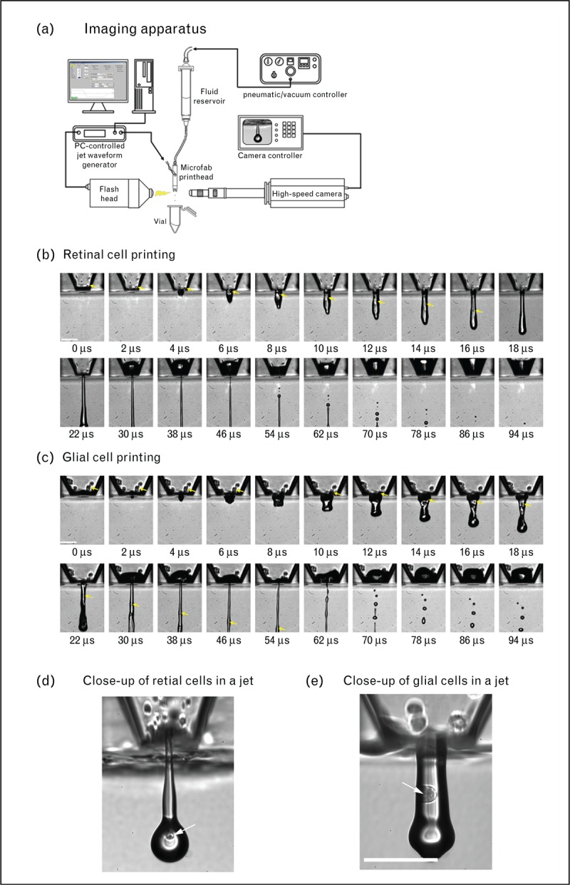
FIGURE 1.

(a) Schematic of an inkjet printing and imaging apparatus that was used to print purified retinal glial and dissociated retinal cells. Image sequences of (b) retinal cells and (c) purified glial cells as they were ejected from the nozzle. The arrows indicate individual cells. Close-up images of (d) retinal cells and (e) glial cells. Scale bar: 100 μm. Reprinted with permission from [10▪].
