
Fig. 6.
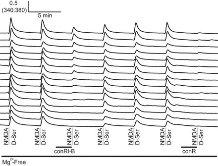
Cerebellar neurons expressed NR2A and NR2B NMDA receptor subtypes in a spectrum of ratios. The combined use of conRl-B and conantokin R (conR) revealed different expression levels for NR2A and NR2B in cerebellar neurons. Cells that were weakly affected by 3.3 μM conRl-B, yet completely inhibited by 3.3 μM conR, are shown at the top and represent cells that express a mixture of NR2B and NR2A NMDA receptors. Cells strongly inhibited by 3.3 μM conRl-B and 3.3 μM conR are shown at the bottom, representing cells that predominantly express NR2B.