Fig. 7.

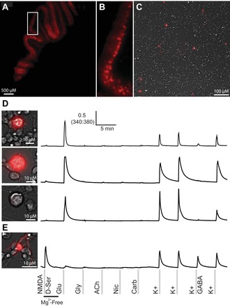
Reporter mice, with genetically labeled neurons expressing tdTomato, facilitated identification of Purkinje neurons in culture. Red fluorescence of the Purkinje cell monolayer from a postnatal day 12 (P12) mouse is shown (A and B). Fluorescent neurons in a culture of cerebellar cells (overlaid on a brightfield image) from a P7 mouse are shown (C). D: three different Purkinje neurons and their response profiles. Purkinje neurons were large and round, brightly fluorescent, and responded to glutamate and depolarization. However, not all Purkinje neurons were labeled at P7. The genetic labeling of Purkinje neurons has been reported to be complete by P21. This also shows a large and round nonfluorescent cell with the same profile as the fluorescent cells. E: a small fluorescent cell with a different morphology in culture and a different response profile that was consistent with granule cells.
