
Fig. 8.
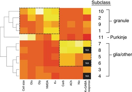
Heat map of mean values for 11 cerebellar cellular subclasses presented in Table 1. The color scale varies from red (low) to yellow (high). For cell size, yellow represents an average large cell area and red, a small cell area. For each pharmacological challenge, yellow represents an average large response and red, a small response. Black represents data that are not available (NA), because the corresponding cells did not respond to K+ depolarization. Hence, their K+ response could not be blocked by GABA. Cellular subclasses have been clustered and ordered according to the similarities in mean values for each variable. Subclasses 1 and 2, proposed to be granule cells, cluster with subclasses 9 and 10, which may represent additional granule cell subclasses. Subclass 11 is comprised of the Purkinje neurons that were identified by genetic labeling from reporter mice, cell morphology, and a distinctive functional profile. Additional cellular subclasses that did not respond to depolarization (4, 6, and 8) are likely to be glial cells and have clustered with other acetylcholine-responsive cellular subclasses at the bottom of the heat map. The categorization of subclasses 3, 5, and 7 as neuronal or glial cells is ambiguous, because they responded relatively weakly to depolarization.