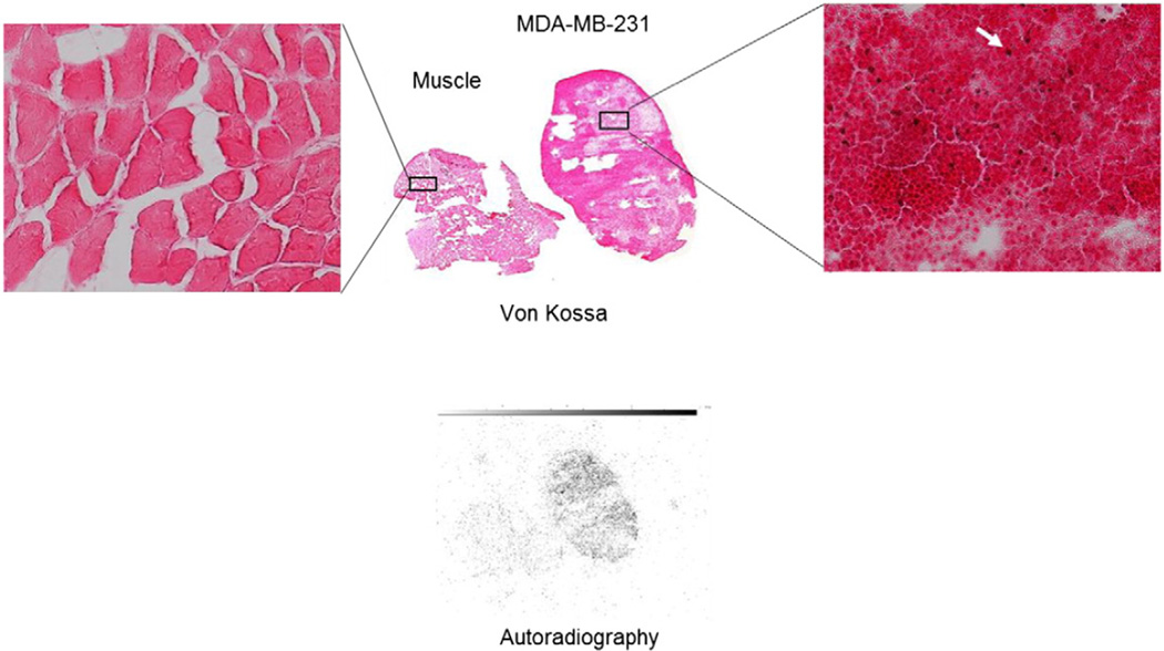
Fig. 3.
Von Kossa (colored) and autoradiography slices (black and white) of muscle and an MDA-MB-231 tumor harvested from a mouse immediately following SPECT imaging with 99mTc-MDP. The far left and far right inserts are magnified images of the stained samples. The red/brown von Kossa stains (pointed to by the white arrow) indicate the presence of type II microcalcification. Distribution of 99mTc-MDP was well correlated with the positive von Kossa stains while no autoradiography signal or positive stains were found in the muscle tissue.

