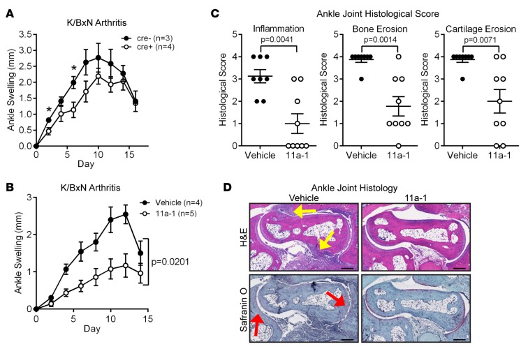
Figure 6. In vivo inhibition of SHP-2 attenuates arthritis in mice.
(A) At 6 to 10 weeks of age, female Ptpn11fl/+-Mx1 cre– (n = 3) and cre+ (n = 4) mice were injected i.p. with 3 doses of 300 μg polyinosinic-polycytidylic acid [poly(I:C)] every other day. Ten days after the last poly(I:C) treatment, mice were injected i.p. with 200 μl K/BxN sera to induce arthritis. Ankle thickness was measured every other day after serum transfer. Mean ± SEM ankle swelling is shown. *P < 0.05, 1-tailed unpaired t test. (B–D) Female C57BL/6 mice were administered 200 μl K/BxN sera at 8 weeks of age and administered 7.5 mg/kg 11a-1 (n = 5) or vehicle (n = 4) i.p. daily for 14 days. (B) Mean ± SEM ankle swelling is shown. Data were analyzed using 2-way ANOVA. (C and D) Histological analyses of ankles stained with H&E or Safranin-O at the end of the disease course. (C) Scatter plots show the mean ± SEM histological score of each ankle. Data were analyzed using the 2-tailed Mann-Whitney test. (D) Representative images of H&E-stained (yellow arrows indicate regions of inflammatory infiltrate) or Safranin-O–stained (red arrows indicate regions of cartilage erosion) joints are shown. Scale bars: 200 μm.

