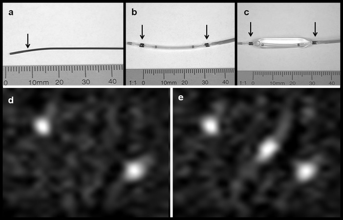
Fig 4. Photographs and MPI of guidewire and PTA balloon catheter.
(a) Photography of the magnetically labeled RadiofocusFM guidewire (arrow indicates the end thin lacquer-labeling of the tip). (b) Photography of the deflated and (c) with saline inflated balloon catheter with magnetic markers at both balloon ends (arrows). (d) MPI-maximum intensity projection (MIP) in z-projection of the balloon catheter showing the two marks as bright spots. (e) MPI-MIP in z-projection of the balloon catheter coaxially placed on the guide wire with the guidewire tip at its center.

