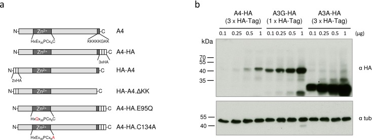
Fig 2. Expression of the A4-HA fusion proteins.
(a) Schematic representation of protein domains and motifs found in the human A4 protein and tested variants. Zn2+: presumed zinc-binding domain. HA (white boxes): HA-tag. KKKKKGKK: polylysine domain. (b) Increasing amounts of A4-HA (3xHA-tags), A3G-HA (1xHA-tag) and A3A-HA (3xHA-tags) expression plasmids were transfected into 293T cells followed by immunoblot analysis of the transfected cells using an anti-HA antibody. Immunoblot analysis with anti-tubulin (tub) antibody served as loading control. α, anti.

