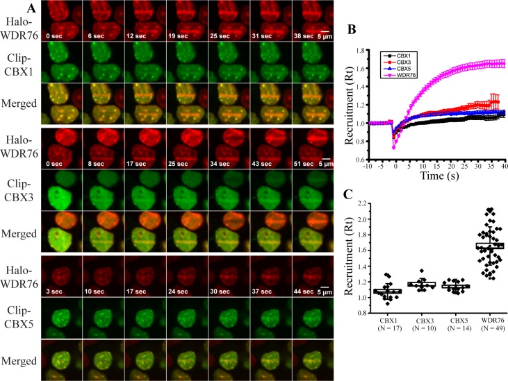
Fig 5. Co-localization of WDR76 with Heterochromatin Proteins.
(A) HEK293FRT cells stably expressing Halo-WDR76 and transiently expressing CLIP-CBX1, CLIP-CBX3, and CLIP-CBX5 were stained with Hoechst dye, micro-irradiated with a 405 nm UV laser. Merged images of both labeled proteins after laser microirradiation are also shown. In cases where there is more than one cell in image the two cells not damaged simultaneously. (B) Kinetics of recruitment to microirradiated regions of WDR76 (pink), CBX1 (black), CBX3 (red), and CBX5 (blue). Cells were imaged every second, and intensity values were binned over 5-s intervals. Microirradiation was initiated at time = 0s. Averages are shown with the standard error. (C) Kinetics of recruitment to microirradiated regions of WDR76, CBX1 and CBX3 represented as box plots with the box showing the standard error with a coefficient of 1 and the whiskers showing the standard deviation with a coefficient of 1.

