
Figure 1.
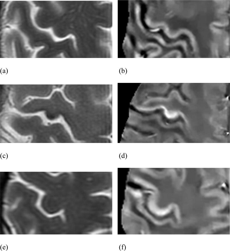
Axial T2-weighted and QSM images in a 50-year-old male control patient (a and b); a 42-year-old man with definite ALS (c and d), with an MRC upper extremity strength score of 57/100 and an ALS-FRS-R of 30; and a 49-year-old woman with probable ALS (e and f), with an MRC upper extremity strength score of 82/100 and an ALS-FRS-R of 43. Of note, the patient with definite ALS in Figs. 1c and 1d presented with profound left upper extremity weakness, and the RMCS scores for his right and left hand lobules were 89.3 ppb and 74.2 ppb, respectively.