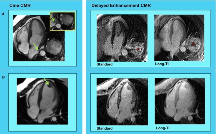
Figure 1.

Cardiac metastasis (CMET) morphology and tissue properties. Representative examples of CMETs as assessed by cardiac magnetic resonance (CMR) imaging. A, Irregularly contoured left atrial mass (green arrow) in a patient with a testicular germ cell tumor. Cine‐CMR (left) demonstrates direct extension via the left lower pulmonary vein. Delayed enhancement (DE‐)CMR tissue characterization (right) demonstrates heterogeneous enhancement, including peripheral contrast uptake and central hypoenhancement (asterisk). B, Ovoid left ventricular apical mass (green arrow) in a patient with sarcoma. Note that whereas location and morphology on cine‐CMR (left) suggest thrombus, DE‐CMR tissue characterization (right) demonstrates diffuse contrast uptake—consistent with vascular supply secondary to neoplastic etiology.
