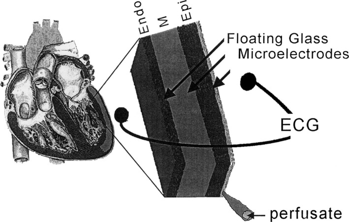
Figure 1.

Arterially perfused left ventricular wedge model of canine myocardium. Schematic diagram of the arterially perfused canine LV wedge preparation. The wedge is perfused by a small native branch of the left descending coronary artery and stimulated from the endocardial surface. Transmembrane action potentials are recorded simultaneously from epicardial (Epi), M region (M), and endocardial (Endo) sites using three floating microelectrodes. A transmural ECG is recorded along the same transmural axis across the bath, registering the entire field of the wedge. Reproduced with permission from Yan et al.9 Promotional and commercial use of the material in print, digital, or mobile device format is prohibited without the permission from the publisher Wolters Kluwer Health. ECG indicates electrocardiogram; LV, left ventricular.
