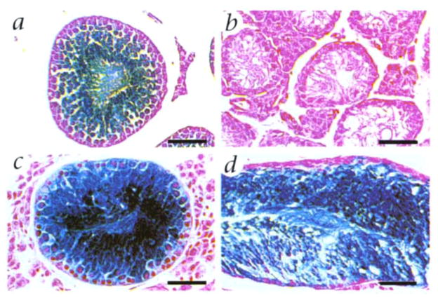
Fig. 2.
Microscopic appearance of spermatogenesis in recipient seminiferous tubules following microinjection of donor testis cells preserved at −196 °C. a, Seminiferous tubule of donor testis from transgenic ZFlacZ mouse. Round spermatids and more mature stages stain blue following incubation with X-gal (Fig. 1a)3,4,11. When staining is intense, immature stages also appear blue. b, Seminiferous tubule from recipient mouse treated with busulfan. No germ cell stages are present. Only Sertoli cells remain. c and d, Seminiferous tubules from busul-fan-treated recipient mice 891 and 771, respectively (Table 1). Blue staining of germ cells indicates their origin from transplanted donor cells that had been frozen 7 and 111 days, respectively. Background stain in all sections is neutral fast red. Scale bar, 50 μm.

