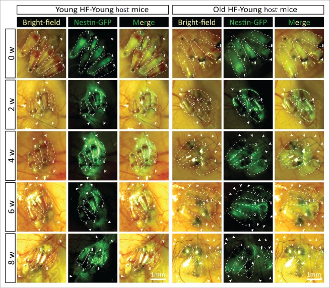
Figure 4.
Time-course comparison of ND-GFP fluorescence of HAP stem cells and their location in young and old hair follicles transplanted to young nude host mice. ND-GFP expressing HAP stem cells were located in various areas: sensory nerve, hair matrix bulb area, and outer-root sheath area. HAP stem cell ND-GFP fluorescence was imaged with the Dino-Lite. In the fluorescence images, each follicle is outlined with a dashed line and numbered for comparison with the brightfield images where the follicles are numbered. Please see the Materials and Methods for details.

