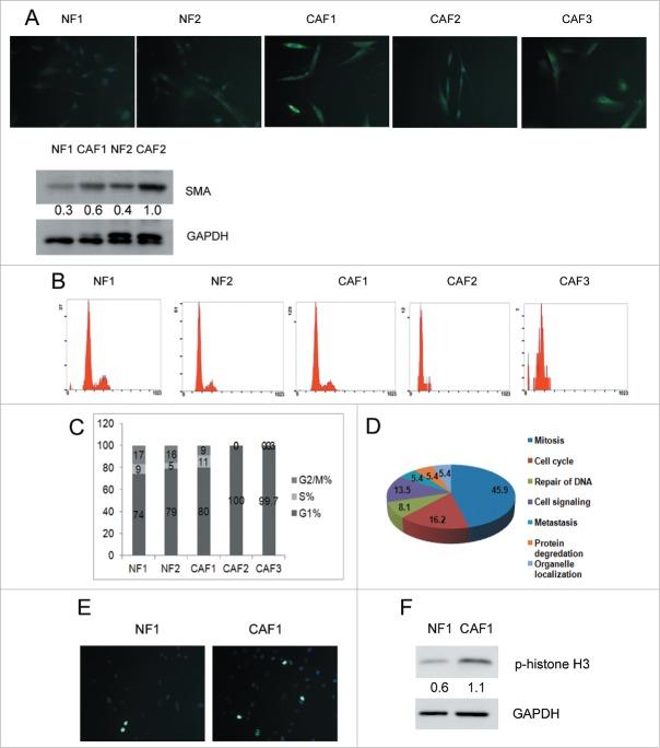
Figure 1.
CAF1 cells are mitotically active. (A) Identification of human primary ovarian NFs and CAFs. Immunostaining of FSP1 and western blot analysis of α-SMA in NFs and CAFs. Cells were stained with FSP1 (green) and counterstained with DAPI (blue). The relative intensity of α-SMA normalized to housekeeping protein was shown at the bottom. (B) Cell cycle analysis of NFs and CAFs. (C) The cell cycle phase distribution in each cell line (as in B). (D) Pathways enriched in CAF1 was revealed by GSEA indicated that CAF1 cells were mitotically active. (E) Immunostaining of phospho-histone H3 in NF1 and CAF1 cells. Cells were stained with phospho-histone H3 (green) and counterstained with DAPI (blue). (F) Western blot analysis of phospho-histone H3 in NF1 and CAF1 cells. The relative intensity of phospho-histone H3 normalized to housekeeping protein was shown at the bottom.

