Figure 3.
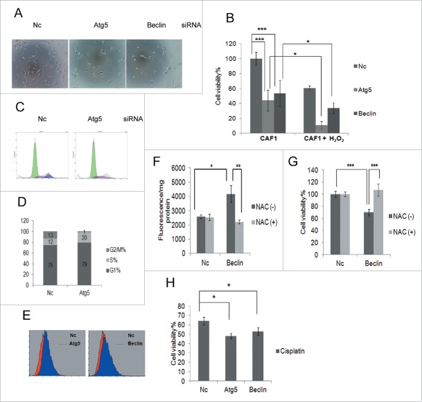
Autophagy protects ovarian CAF1 cells against oxidative stress by inhibiting ROS production. (A) The representative images of CAF1 cells with knockdown of indicated siRNA and treated with H2O2. Nc: negative control siRNA. (B) The effect of blockage of autophagy on cell viability and H2O2-induced cell death of CAF1 cells. CAF1 cells were reverse transfected with the indicated siRNA for 48 h in 96-well plate in triplicate, treated with or without 1 mM H2O2 for 2 h. The cell viability was measured with MTT assay (mean ± SD, n = 3). (C) Cell cycle analysis of CAF1 cells transfected with Nc or Atg5 siRNA for 48 h. (D) The cell cycle phase distribution in CAF1 cells transfected with Nc or Atg5 siRNA for 48 h (as in C). (E) ROS measurement of CAF1 cells transfected with Nc, Atg5 or Beclin siRNA for 48 h by flow cytometry. (F) NAC decreased ROS production in Beclin knockdown CAF1 cells. Cells were transfected with Nc or Beclin siRNA for 24 h, incubated with or without NAC (5 mg/ml) for 2 h, and further cultured in complete media for another 24 h. Cells were stained with H2DCF (20 μM) and lysed with RIPA buffer. The fluorescence was read with a fluorometer and normalized to protein concentration (mean ± SD, n = 4). (G) NAC restored cell viability of Beclin knockdown CAF1 cells. Cells were prepared the same way as in (F). Cell viability was measured by MTT assay (mean ± SD, n = 4). (H) The effect of blockage of autophagy on cisplatin-induced cell death of CAFs. CAF1 cells were reverse transfected with the indicated siRNA for 24 h in 96-well plate in triplicate, treated with 100 μM cisplatin for another 24 h. The Cell viability was measured with MTT assay (mean ± SD, n = 3).

