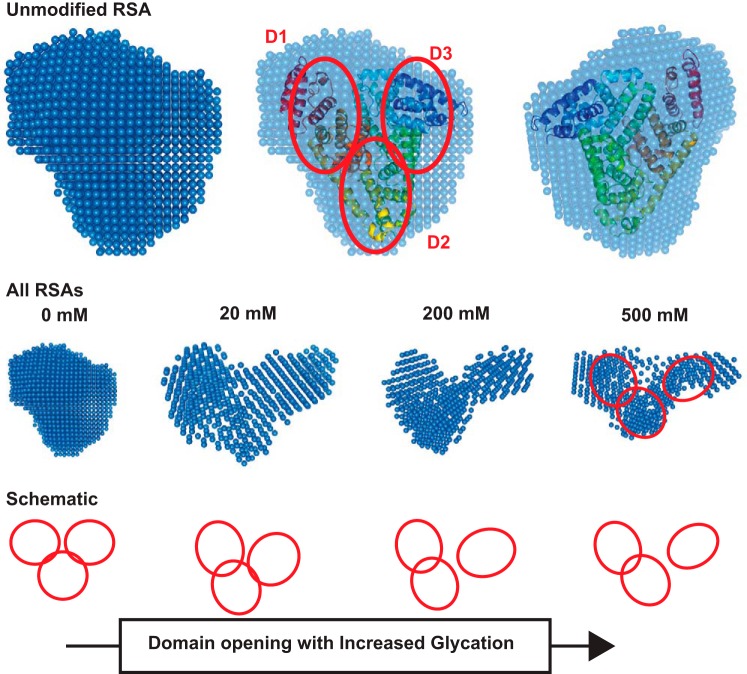
Fig. 6.
SAXS density models of RSA. Top: SAXS data-based uniform density model of unglycated RSA, its overlay with the crystal structure of ligand-free human serum albumin (Protein Database ID 4K2C), and an orthogonal view of the overlaid SAXS envelope. The red ellipses show the domains of albumin. Middle: average uniform density models of glycated versions of RSA. Bottom: schematic representation how domain opening and movement lead to shape changes upon increasing glycation.

