. 2016 Jun 1;5:e13325. doi: 10.7554/eLife.13325

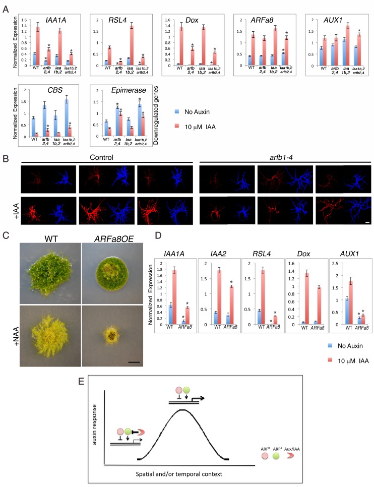
Figure 5. Both repressing and activating ARFs affect auxin response in the same direction in the presence of the Aux/IAAs.

(A) qPCR showing the expression levels of auxin responsive genes in WT, arfb2 arfb4Δ (arfb2,4), iaa1b iaa2Δ (iaa1b,2), and arfb2 arfb4 iaa1b iaa2Δ (iaa1b,2 arfb2,4), mock- or 10 μM IAA-treated for five hours. Error bars represent s.e.m. *p<0.05 (t-test comparing the arfb2 arfb4Δ, and arfb2 arfb4 iaa1b iaa2Δ lines to either WT or iaa1b iaa2Δ, respectively), n=3. (B) Three independent arfb1-4 RNAi and control lines mock- or 10 μM IAA-treated. Each line is presented by two images (DsRED fluorescence (red), chlorophyll auto-florescence (blue). Each image is from projection stacks of multiple confocal sections. Scale bar: 100 μm. (C) WT and an ARFa8 overexpression line (ARFa8OE) grown for one month on BCDAT without auxin or with 12.5 μM NAA. Scale bar: 0.5 cm. (D) qPCR showing the expression levels of auxin responsive genes in WT and ARFa8OE mock- or 10 μM IAA-treated for five hours. Error bars represent s.e.m. *p<0.05 (t-test comparing the ARFa8OE line to WT), n=3. (E) Model for auxin regulation of transcription. The expression level of an auxin responsive gene is determined by the interplay between the Aux/IAAs, the repressing ARFs, and the activating ARFs. At low auxin levels the Aux/IAAs provide stringent repression. At high auxin levels, the Aux/IAAs are degraded resulting in increased transcription through the action of the activating ARFs. The repressing ARFs act to buffer gene expression by attenuating the activity of the activating ARFs. The interplay between the three protein groups results in a wide range of gene expression levels that contribute to a dynamic and context specific auxin response.

DOI: http://dx.doi.org/10.7554/eLife.13325.014

Figure 5.

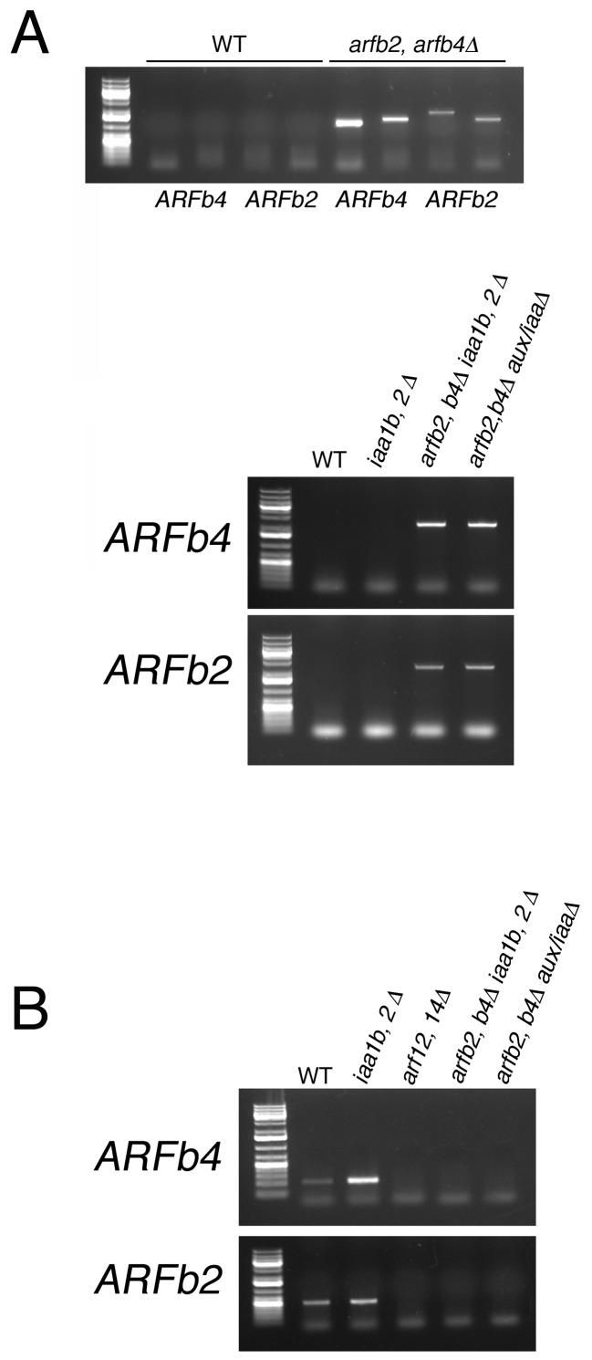
Figure 5—figure supplement 1. PCR detecting the insertion position of different arfb2 arfb4Δ mutants.

Figure 5—figure supplement 1.

(A) Validating the absence of the genomic sequence of ARFb2 and ARFb4PCR. Upper panel: validating the transgene integration in arfb2 arfb4Δ mutant. Primer pairs used to confirm the presence of either up or downstream transgene-endogenous sequence junctions, respectively: ARFb2 (PML703, 722; PML267, 704), ARFb4 (PML195, 675; PML197, 676). Lower panel: validating the transgene integration in arfb2 arfb4Δ iaa1b iaa2Δ and arfb2 arfb4Δ aux/iaaΔ mutants after CRE/lox recombination. Primer pairs used to confirm the absence of the endogenous sequences: ARFb2 (PML703, 704), ARFb4 (PML675, 676). WT sequences were not amplified using these endogenous primers since the resulting fragments are too long and cannot be amplified under the PCR conditions used. (B) Validating the absence of the coding region of ARFb2 and ARFb4 in cDNA samples by RT-PCR. Primer pairs used: ARFb2 (PML551, 612) ARFb4 (PML512, 513).
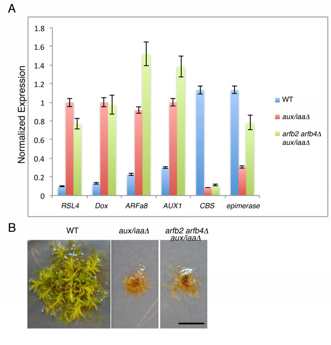
Figure 5—figure supplement 2. Arfb2 and arfb4 Knockout in aux/iaaΔ mutant background (arfb2 arfb4Δ aux/iaaΔ) does not result in phenotypes comparable to reduced auxin sensitivity.

Figure 5—figure supplement 2.

(A) qPCR showing the expression levels of auxin responsive genes in WT, aux/iaaΔ, and arfb2 arfb4Δ aux/iaaΔ. (B) arfb2 arfb4Δ aux/iaaΔ and aux/iaaΔ mutants grown for one month on BCD. Scale bar: 0.5 cm.
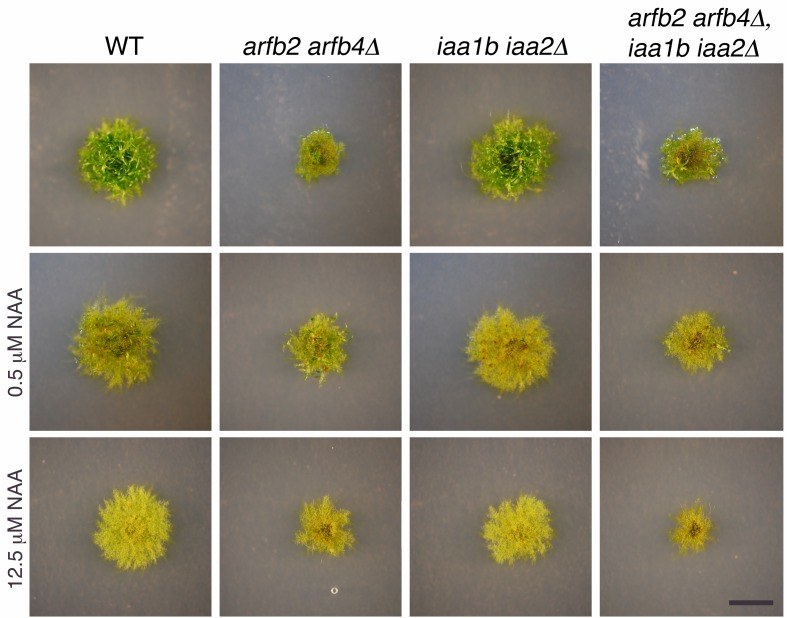
Figure 5—figure supplement 3. Phenotypic analysis of arfb2 arfb4Δ in WT and iaa1b iaa2Δ mutant background.

Figure 5—figure supplement 3.

WT, arfb2 arfb4Δ, iaa1b iaa2Δ, and arfb2 arfb4Δ iaa1b iaa2Δ lines grown for one month on BCDAT medium without auxin or with different NAA concentrations. Scale bar: 0.5 cm.