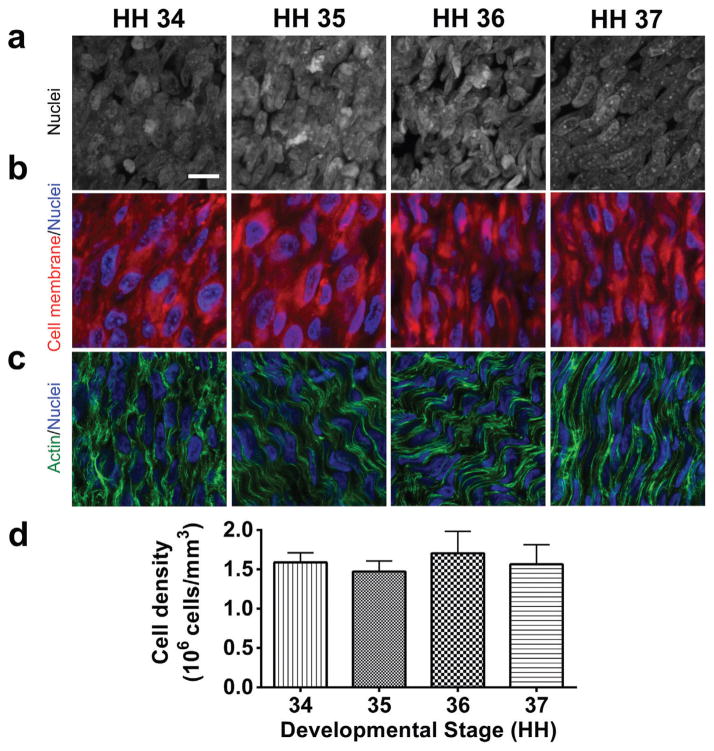
Figure 1.
HH 34–37 embryonic tendons have high cell density throughout early stages of tendon development, as seen by (a) representative 2-dimensional projections of z-stack confocal images of DAPI-stained cell nuclei in whole mount embryonic tendons, and (b) CellMask Plasma Membrane staining (red) for cell membranes, and DAPI staining (blue) for cell nuclei. (c) Phalloidin (green) and DAPI (blue) staining showed organized actin filaments formed an apparent contiguous network spanning adjacent cells (bar, 10 μm). (d) Cell density quantified with image analysis was constant between stages.

