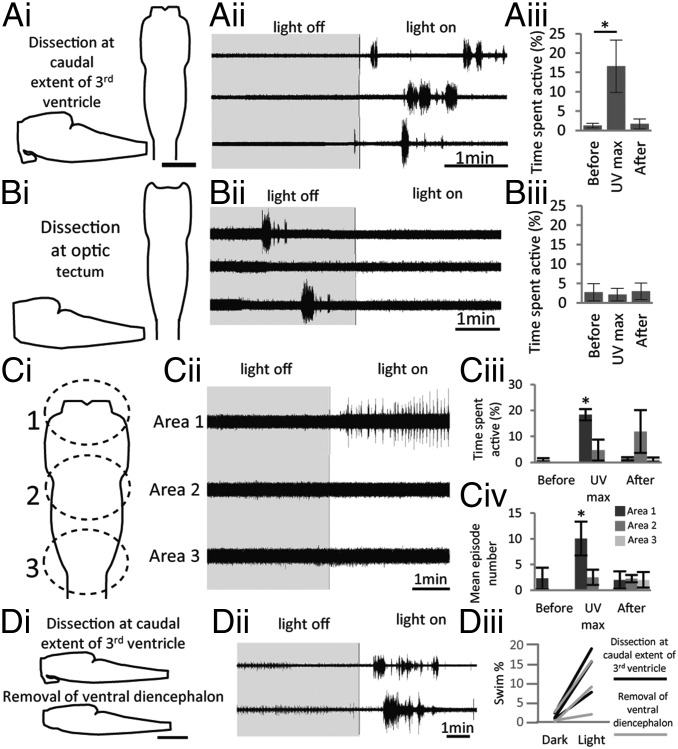
Fig. 3.
Photosensitive tissue resides within the caudal diencephalon. (A, i) Schematic of the dissection performed: The forebrain is removed apart from a small portion of diencephalon caudal to the dorsal opening of the third ventricle. Both dorsal and sagittal aspects are depicted. (Scale bar, 200 µm.) (A, ii) Ventral root recording from a stage 54 larva showing three consecutive responses to illumination with UV light (400 nm; 39 lx). (A, iii) Graph of average data comparing time spent active 200 s before, during, and after illumination (n = 7). (B, i) Schematic illustrating the dissection made flush with the optic tectum to completely remove the diencephalon. (B, ii and iii) Equivalent data to A displayed for preparations lacking the diencephalon (n = 4). (C, i) Schematic illustrating the approximate location of focal illumination of three areas of isolated nervous system. (C, ii) Sequence of responses to illumination of these different areas with UV light following 10 min in the dark (gray box). (C, iii and iv) Graphs showing average data for time spent active (C, iii) and mean episode number (C, iv) for illumination of each area; comparison of 200 s before, during, and after illumination are plotted (n = 4). (D, i) Schematic illustrating isolated nervous system before (Upper) and after (Lower) removal of the ventral diencephalon containing the hypothalamus and pituitary. (D, ii) Ventral root recording from a stage 56 larva before (Upper) and after (Lower) the dissection was performed. (D, iii) Graph illustrating data from three different preparations. Swim % is shown both before (black line) and after (gray lines) removal of the ventral diencephalon. Error bars represent ±SEM; *P < 0.05.

