Fig. S3.
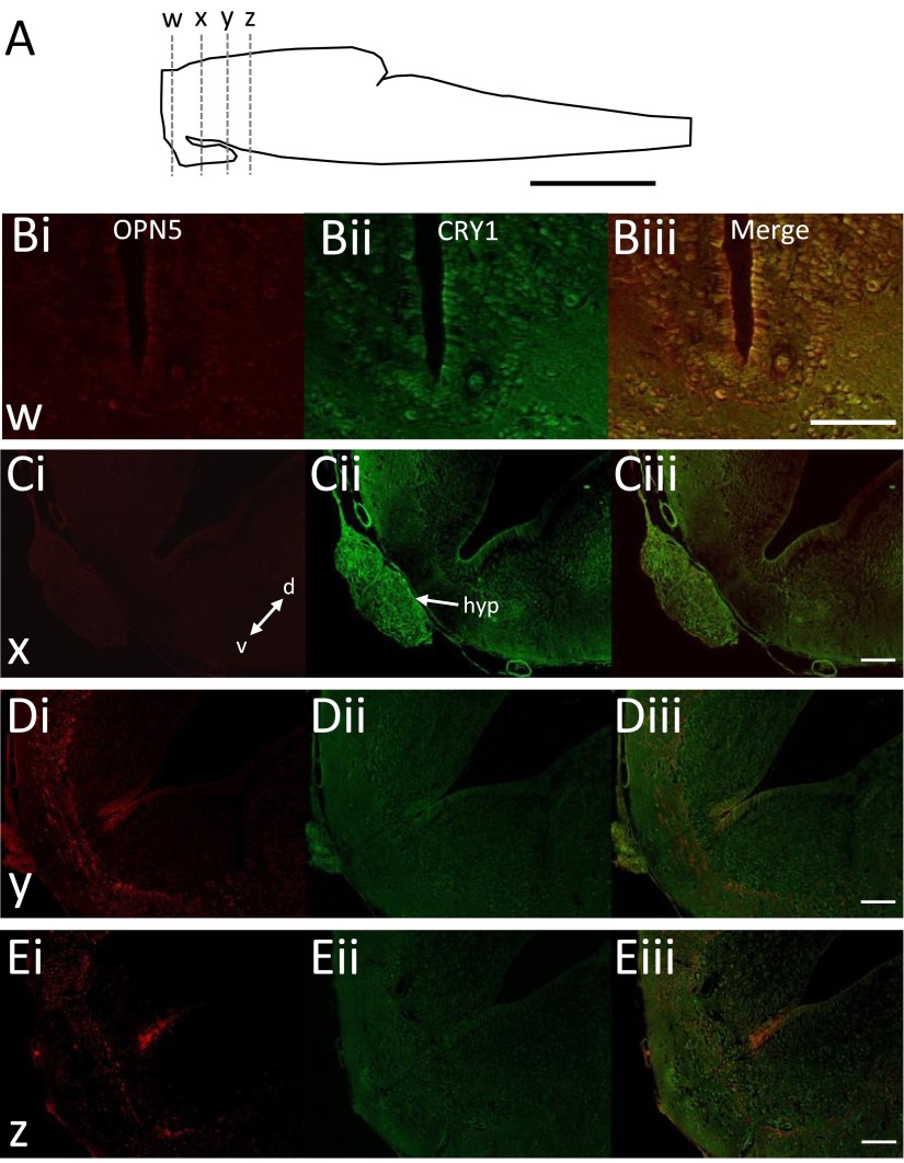
OPN5 and CRY1 are expressed throughout the nervous system. (A) Schematic of a Xenopus tadpole brain showing the approximate position of sections taken for imaging. (B–E) Immunohistochemistry showing OPN5 and CRY1 expression within the ventral half of the preparation at three distinct anatomical levels moving caudally from the region of light sensitivity. d, dorsal; v, ventral. (C, ii) CRY1 is strongly expressed within the hypothalamus (hyp), the region surgically removed in Fig. 3D, without loss of light sensitivity. [Scale bars, 200 μm (A) and 100 μm (B–E).]

