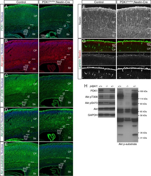
Fig. S1.
Characterization of the brain of PDK1flox/flox;Nestin-Cre mice. (A–G) Immunohistofluorescence analysis of coronal sections of the brain of control or PDK1flox/flox;Nestin-Cre mice at P0 with antibodies to the indicated proteins. PDK1 is expressed ubiquitously in the brain of control mice (A), being most abundant within the CP, where neurons reside. PDK1 expression was efficiently attenuated in the brain of PDK1flox/flox;Nestin-Cre mice (A), but the intensity of Akt (pan) staining within the neocortex was similar for both genotypes (B). Phosphorylation of Akt at Thr308 (T308) is essential for its activation and is mediated by PDK1, whereas phosphorylation at Ser473 (S473) mediated by the mTORC2 complex is thought to modulate Akt function. Phosphorylation of Akt at Thr308 was detected throughout the neocortical wall, most prominently within the CP, of the control brain, but was efficiently attenuated in the PDK1flox/flox;Nestin-Cre neocortex (C). In contrast, phosphorylation of Akt at Ser473 was similar in the control and mutant brains (D). Monitoring of the kinase activity of Akt with antibodies to phosphorylated Akt substrates that harbor a typical Akt phosphorylation consensus sequence revealed that such activity was apparent throughout the neocortex of control mice, being most evident within the CP (E), similar to the distribution of Thr308-phosphorylated Akt (C). In contrast, PDK1 knockout markedly reduced the level of Akt kinase activity (E), although Akt activation remained evident in subplate cells, likely as a result of inefficient PDK1 ablation achieved in this cell population with the use of our Nestin-Cre line (A, C, and E). Examination of the radial glial scaffold, basal lamina, and Cajal–Retzius cells with antibodies to Nestin, to laminin, or to Reelin, respectively, revealed no discernible differences between the two genotypes (F and G). Nuclei were stained with Hoechst 33342. MZ, marginal zone, Str, striatum. (Scale bar, 100 µm.) (H) Immunoblot analysis of the neocortex of mice of the indicated conditional Pdpk1 genotypes at P0 confirmed that Pdpk1 ablation resulted in loss of PDK1 protein and attenuation of the phosphorylation of both Akt at Thr308 and Akt substrates. GAPDH was examined as a loading control.

