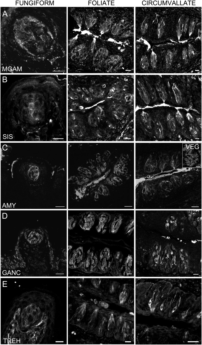
Fig. 3.
Expression of α-glucosidase proteins in taste cells. Indirect immunofluorescence confocal microscopy of taste bud containing sections from mouse FNG, FOL, and CV taste papillae was carried out with specific polyclonal antibodies directed against AMY1/2, MGAM, SIS, GANC, and TREH. Immunofluorescence indicates expression in taste cells of all five enzymes. [Scale bars, A = 10 μm (FNG), 40 μm (FOL and CV); B = 40 μm (all); C = 80 μm (FNG), 20 μm (FOL and CV), and 40 μm (VEG); D = 80 μm (FNG), 40 μm (FOL and CV); E = 10 μm (FNG), 20 μm (FOL and CV).]

