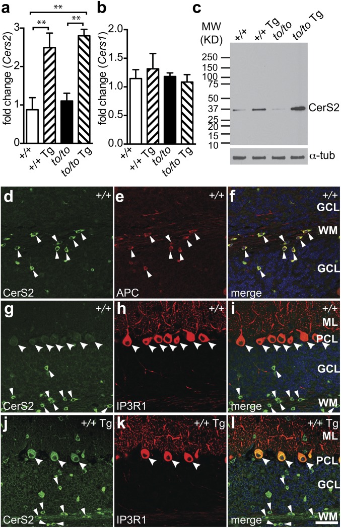
Fig. 1.
Cerebellar neuronal expression of CerS2 from a transgene. (A and B) Real-time PCR results showing changes of the expression of Cers2 (A) and Cers1 (B) transcripts in brains of 4-wk-old Cers1 wild-type (+/+), Cers1 wild-type with the neuron-specific Cers2 transgene (+/+ Tg), homozygous Cers1 toppler mutant (to/to), and Cers1 toppler mutant with the Cers2 transgene (to/to Tg). Values are mean ± SD (three mouse samples, four technical replicates for each sample). **P ≤ 0.01 (one-way ANOVA, multiple comparisons). (C) A representative Western blot analysis showing the CerS2 protein level in the brain. Genotypes of mice are as described in A and B. The blot was redeveloped with an antibody against α-tubulin as a loading control. (D–l) Immunohistochemistry of brain sections from 4-wk-old wild-type (D–I, +/+) and Tg-CerS2 (J–L, +/+ Tg) mice, using antibodies against CerS2 (D, G, and J), APC (E), and IP3R (H and K). Merged images are also shown (F, I, and L). Purkinje cells and oligodendrocytes are marked with large and small arrowheads, respectively. Cerebellar cell layers are labeled as GCL (granule cell layer), WM (white matter), ML (molecular layer), or PCL (Purkinje cell layer). (Scale bar, 50 µm.)

