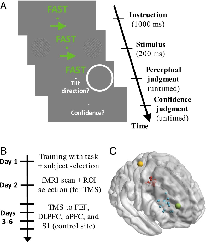
Fig. 1.
Task, experiment time line, and TMS locations. (A) Trial sequence. Each trial began with a 1-s instruction to attend to either the left or right stimulus, as well as to emphasize either speed or accuracy. The grating stimuli were presented for 200 ms, and a postcue indicated which stimulus subjects should respond to. The postcue was on the attended side 66.7% of the time. Responses regarding stimulus orientation (clockwise/counterclockwise) and confidence (on a 1–4 scale) were untimed. The following trial began 1 s later. (B) Experiment time line. ROI, region of interest. (C) Approximate location of S1 is depicted in yellow (the target was identified in the postcentral gyrus). FEF (red) and DLPFC (blue) were localized separately for each subject based on individual fMRI activations (each dot represents a different subject). Finally, the site for aPFC stimulation (green) was common across subjects and based on Fleming et al. (4). All targets were identified in the right hemisphere. The y coordinates for each region did not overlap: S1: −33, FEF: [−10, 2], DLPFC: [26, 48], aPFC: 53.

