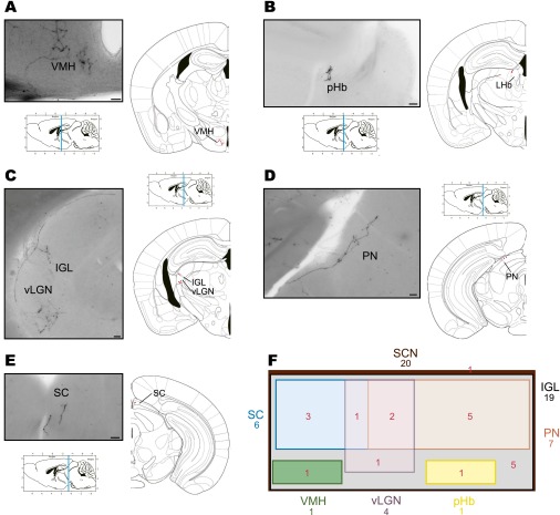
Fig. S2.
A single M1 ipRGC innervates multiple brain targets. Representative images of brain targets from a single ipRGC in the mouse are shown in A–E. Single ipRGCs innervate the SCN and send collateral projections to the ventral medial hypothalamus (VMH) (A), peri-Habenular region (pHb) (B), intergeniculate leaflet (IGL) and ventral part of the lateral geniculate nucleus (vLGN) (C), pretectal nucleus (PN) (D), and superior colliculus (SC) (E). (F) Venn diagram showing the overlap between brain nuclei innervated from 20 individual ipRGCs. The surface is proportional to the percentage of ipRGC innervation to brain targets. (Scale bars: 50 μm.)

