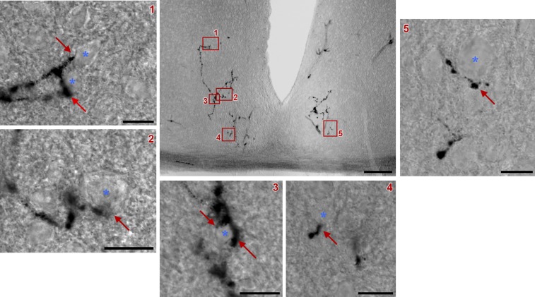
Fig. S4.
ipRGC axonal terminals within the SCN. A representative coronal section of the SCN after AP staining is shown (Scale bar: 100 μm.) Retinal fibers from a single ipRGC (arrows) are in close contact with neurons (asterisks) located in different SCN regions (Scale bars: 10 μm.) In most cases, clear terminal swellings were observed. Red boxes 1–5 from the central image are magnified for better viewing.

