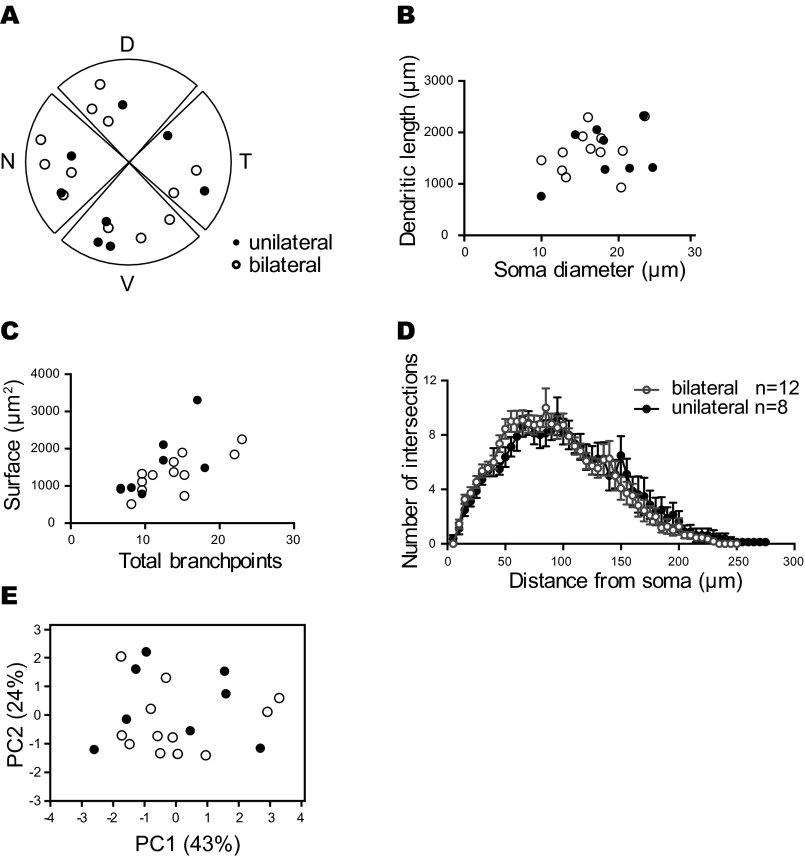
Fig. S6.
Unilateral and bilateral projecting ipRGCs have similar somato-dendritic features. (A) Composed spatial distribution of the cell bodies from unilateral (closed circle) and bilateral (open circle) projecting ipRGCs. Although unilateral and bilateral ipRGCs showed distinct axonal architectures in the SCN, they were similarly distributed in the retina. (B–E) Dendritic morphology of unilateral (closed circle) and bilateral (open circle) projecting ipRGCs showed similar properties in x–y scatter plot with soma diameter versus dendritic length (B), total branch points versus dendritic surface (C), and dendritic Sholl analysis (D). In addition, unilateral and contralateral projecting cells are clustered together in the PCA plotted with components 1 (PC1, 43% of explained variance) and 2 (PC2, 24% of explained variance) using all somato-dendritic features from 20 cells (E).

