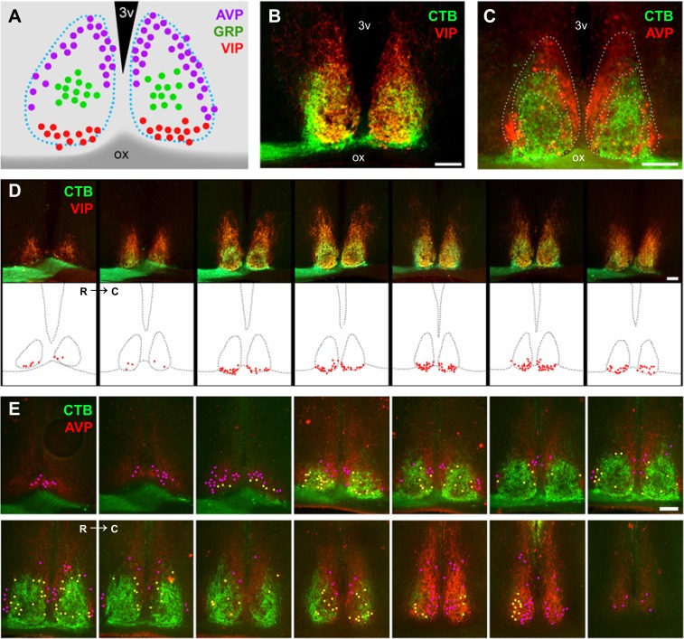
Fig. S9.
Topography of peptidergic neurons in the SCN. (A) VIP, GRP, and AVP cells show a particular topography in the SCN. VIP cells (B and D) are located in the most ventral SCN. Most VIP cells are in close contact with retinal fiber coming from the optic chiasm. AVP cells (C and E) were found in the shell area. Based on retinal projections and the potential innervation, AVP cells were divided into two distinct populations: AVP cells located in the outer nonretinorecipient shell (cells in magenta) and AVP cells located in the internal shell region that receives retinal innervation (cells in yellow). C, caudal; ox, optic chiasm; R, rostral; 3v, third ventricle. (Scale bars: 100 μm.)

