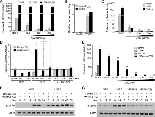
Fig. 3.
USP6 regulates WNT signaling at the level of the Wnt ligand and receptor complex. (A) Inhibition of USP6 induced Wnt signaling by XAV939. HEK293T cells with integrated Wnt/β-catenin reporter were transfected with USP6 expression plasmid. Following treatment with increasing concentrations of XAV939 for 16 h, luciferase activity was measured. β-catenin(SA) serves as a control; it is constitutively active and resistant to degradation by the destruction complex. Data represent mean ± SD. (B) USP6 does not potentiate Wnt/β-catenin signaling induced by GSK3 inhibitor. HEK293T cells stably expressing the Wnt/β-catenin reporter were transfected with control or USP6 expression plasmids. The cells were treated with 1 μM GSK inhibitor CT99021 for 16 h. Luciferase reporter activity as measured is expressed as mean ± SD (n = 3). ns, not significant. (C) Inhibition of USP6-induced Wnt signaling by DKK1. HEK293 cells with an integrated Wnt/β-catenin reporter were transfected with USP6 and/or WNT3A and incubated with varying amounts of DKK1. Data represent mean ± SD (n = 3). (D) LRP6 silencing decreases USP6-induced Wnt signaling. HEK293T cells were transfected with the indicated siRNAs. Forty-eight hours after siRNA transfection with two independent LRP6 siRNAs (siA and siB) or control siRNA (siCNT), plasmids expressing GFP, USP6, or β-catenin(SA) were transfected and cells were treated with control- or WNT3A-conditioned medium. Luciferase reporter activity is represented as mean ± SD (n = 4). (E) C59 inhibits USP6-induced Wnt signaling. HEK293 cells with an integrated Wnt/β-catenin reporter were transfected with USP6 and WNT3A expression plasmids and incubated with the indicated concentrations of C59. Data represent mean ± SD (n = 3). (F and G) USP6 expression increases LRP6 phosphorylation. HEK293T cells were transfected with control or plasmids expressing USP6 or its variants before treatment with control- or WNT3A-conditioned media for the indicated duration. Total cellular LRP6 levels and phosphorylated LRP6 (p-LRP6) levels were determined by Western blot analysis.

