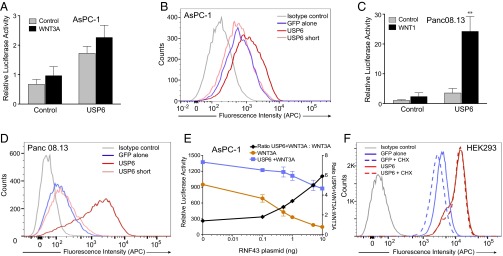
Fig. 5.
USP6 does not potentiate Wnt signaling in RNF43 mutant cells. (A) USP6 does not synergize with WNT3A in RNF43 mutant pancreatic cancer cells. AsPC-1 cells were cotransfected with Wnt/β-catenin reporter and USP6 and WNT3A expression plasmids 24 h before analysis. Relative luciferase activity is plotted; error bars represent SD from the mean (n = 3). (B) Fzd levels in AsPC-1 cells expressing USP6. AsPC-1 cells were transfected with GFP or USP6-GFP and its variants. Cell surface levels of Fzds in GFP-positive cells as analyzed by flow cytometry are shown. Data are representative of three independent experiments. (C) USP6 synergizes in pancreatic cancer cells with WT RNF43. Panc 08.13 cells were transfected as in A. Relative luciferase activity is represented as mean ± SD (n = 3). (D) Fzd levels in Panc 08.13 cells expressing USP6. Panc 08.13 cells were transfected with GFP or USP6-GFP and its variants. Cell surface levels of Fzds in GFP-positive cells as analyzed by flow cytometry are depicted. Data are representative of four independent experiments. (E) Restoration of USP6 synergy in RNF43 mutant cells. AsPC-1 cells were transfected with Wnt/β-catenin reporter and WNT3A and indicated concentrations of RNF43 expression plasmids. Luciferase activity was measured after 24 h. Data represent mean ± SD (n = 3). (F) USP6 prevents down-regulation of Fzd receptors. HEK293 cells were transfected with GFP or USP6-GFP. After 48 h, new protein synthesis was blocked by addition of 40 μg/mL cycloheximide. Ninety minutes later, cell surface levels of Fzds in GFP-positive cells were assessed by flow cytometry. Data are representative of four independent experiments.

