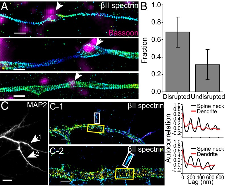
Fig. 4.
The distribution of βII spectrin in axonal boutons and dendritic spines. (A) Representative STORM images of βII spectrin in axons that contain presynaptic boutons marked by a presynaptic marker, Bassoon (magenta). βII spectrin is labeled by expressing GFP-βII spectrin (or GFP-αII spectrin) in a sparse subset of cultured neurons, and hence some bassoon-positive regions do not overlap with the GFP-positive axon. (Top and Middle) Examples of bassoon-positive presynaptic boutons (indicated by arrow) within which the periodic pattern of spectrin is disrupted. (Bottom) An example of bassoon-positive presynaptic bouton (indicated by arrow) within which the period pattern of spectrin is not disrupted. (B) Bar graph depicting relative fraction of boutons in which the periodic structure is disrupted or undisrupted. (C) The dendritic region of a cultured mouse hippocampal neuron immunostained with MAP2, a dendrite marker. (C-1 and C-2) STORM images of two selected regions showing periodic patterns of immunolabeled endogenous βII spectrin in spine necks (Left panels) with autocorrelation functions (Right panels) calculated from the boxed spine neck region (white boxes) and dendritic shaft (yellow boxes). (Scale bar: 10 µm for conventional images and 1 µm for STORM images.)

