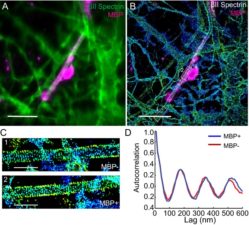
Fig. S6.
In vitro myelination does not affect the formation of the MPS structure. Hippocampal neurons were cultured for 2 wk before adding the glia cells. After coculturing for 6 wk, the cells were fixed and immunostained with βII spectrin (green) and MBP (myelination marker, magenta). (A) Conventional image of neurons showing a myelinated axon (shown in magenta). (Scale bar: 10 µm.) (B) 3D STORM image of βII spectrin in the same area as in A. The myelin sheath was shown in magenta and imaged at the conventional resolution. βII spectrin adopts a highly periodic structure in both the myelinated and unmyelinated regions along the axon. (Scale bar: 10 µm.) (C) Zoom-in 3D STORM image of βII spectrin from the two boxed regions from B). (Scale bar: 2 µm.) (D) Averaged autocorrelation functions of βII-spectrin distribution calculated from multiple MBP+ and MBP− axons (n = 7 each).

