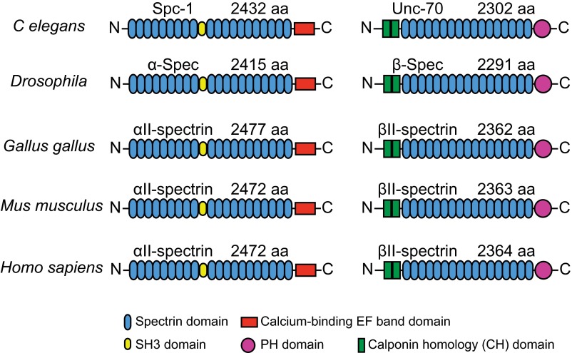
Fig. S8.
Structural domain organization of α and β spectrin across different species. The structural domains of αII and βII spectrin homologs, including spectrin domain (blue), SH3 domain (yellow), calcium-binding EF band domain (red), PH domain (magenta), and calponin homology (CH, green) domain from C. elegans, Drosophila, G. gallus, Mus musculus, and H. sapiens. Also shown are the lengths of these proteins in terms of the number of amino acids (aa).

