
Fig. 6.
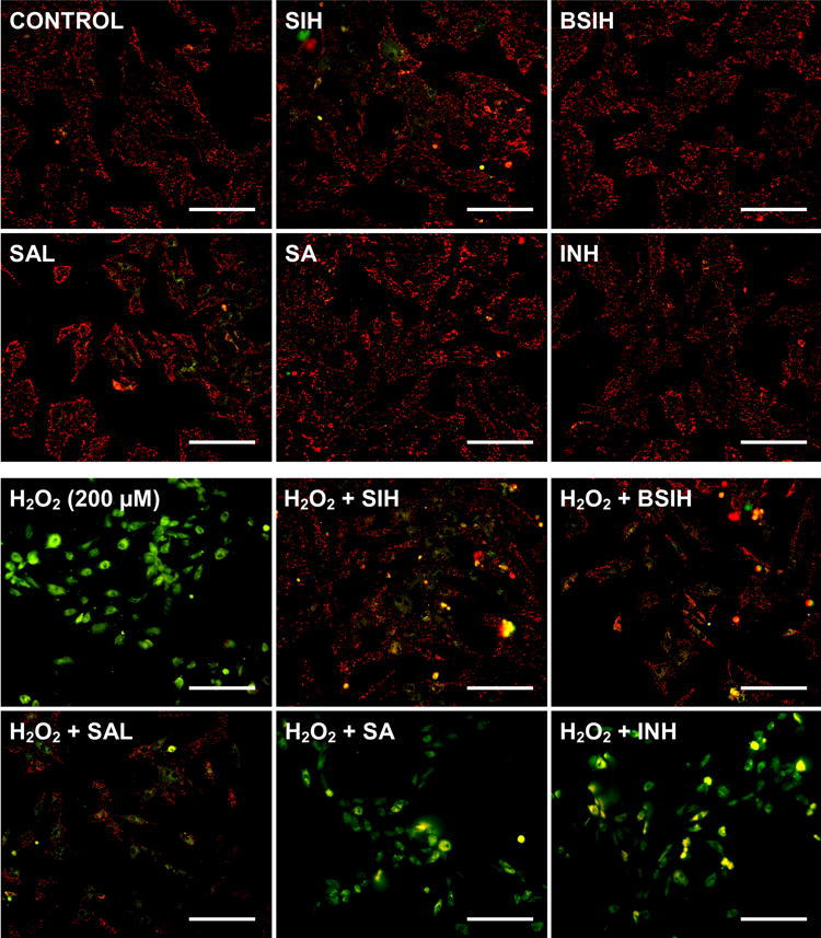
Mitochondrial inner membrane potential (ΔΨm) was assessed by epifluorescence microscopy. H9c2 cells were incubated for 24 h with tested compounds (BSIH, SIH, SAL, SA and INH) or H2O2 (alone or in combinations) and then stained with JC1 probe. Punctate or rod-shaped red emission reflects mitochondrial inner membrane potential-dependent accumulation of probe aggregates in actively respiring mitochondria, diffuse cytosolic green fluorescence indicates monomers of the probe released into cytoplasm after mitochondrial depolarization. Lack of fluorescence is indicative of probe release from necrotic or secondary apoptotic cells. Inherent toxicities of tested compounds (all 100 μM) are shown on the upper photographs and protective effects of these compounds against the dissipation of ΔΨm induced by 200 μM H2O2 are shown on the lower photographs. Scale bars represent 200 μm.