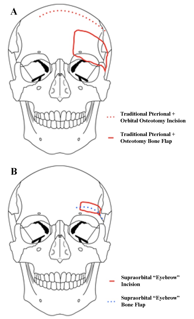
Figure 2. Standard pterional and orbital osteotomy approach (A) vs. supra-orbital approach (B).

In a supraorbital approach, an incision is made within the eyebrow and lateral to the supraorbital nerve, and the frontalis muscle is divided parallel to the orbital rim and reflected downward. The keyhole is made just posterior to the temporal line, and the orbital rim is drilled flush with the orbital roof. Modified from: Rosario Van Tulpe, https://commons.wikimedia.org/wiki/File:SkullSchaedel3.png
