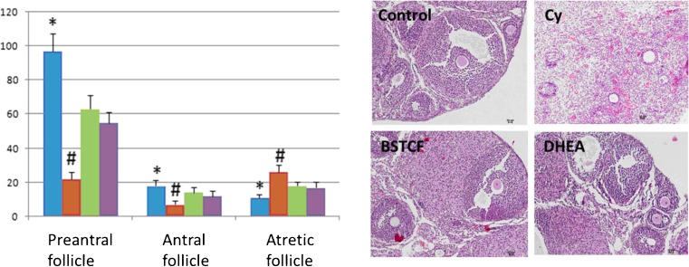
Fig. 1.
Cyclophosphamide injection results in decreased numbers of preantral and antral follicles but increased number of atretic follicles compared to the control (left upper HE staining images) as shown in ovaries (HE staining) which represent DOR (right upper HE staining images). Both BSTCR and DHEA treatments restore these follicle changes (left lower and right lower HE staining images, respectively). All HE staining images were taken at ×10 magnification. Follicles were also quantified by a histogram: the blow column represents the control mice, while the brow column indicates DOR mice; light green column represents mice with BSTCR treatment, while purple column stands for mice with DHEA treatment. The Y-axis represented the number of follicles. *P < 0.05, # P < 0.01

