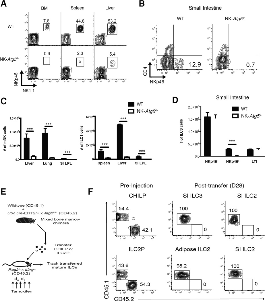
Figure 1. Atg5 is essential for innate lymphocyte development.
(A) Representative flow cytometric plots of lineage marker negative (Lin−) NKp46+NK1.1+ cells and (B) Lin−NK1.1−CD127+CD90.2+Rorγt+ (ILC3s) in indicated peripheral organs of NKp46iCre × Atg5fl/fl mice (“NK-Atg5−/−”) or littermate Atg5fl/fl controls (called “WT”) (C) Absolute numbers of mature NK cells (mNK; Lin−NK1.1+NKp46+DX5+Eomes+), ILC1 (Lin−NK1.1+NKp46+DX5−Eomes−), and (D) ILC3s in indicated peripheral organs of WT or littermate NK-Atg5−/− mice (E,F) WT mice (CD45.1 × CD45.2) were lethally irradiated and reconstituted with an equal number of bone marrow cells from WT (CD45.1) and UbcCre-ERT2 × Atg5fl/fl (CD45.2) mice. (E) Schematic of experiment. (F) Both WT (CD45.1) and UbcCre-ERT2 × Atg5fl/fl (CD45.2) CHILP (Lin−NK1.1−DX5−FLT3−CD127+α4β7+CD25−) and ILC2P (Lin−NK1.1−DX5−FLT3−CD127+α4β7+CD25+) cells were adoptively transferred into sub-lethally irradiated Rag2−/− × Il2rg −/− hosts and injected i.p. with 1mg of tamoxifen daily for 5 days. After 28 days, the SI and adipose tissue were harvested from recipient mice and analyzed for mature ILC2 (Lin−NK1.1−DX5−CD127+CD90.2+GATA3+KLRG1+) and ILC3 cells. (Lin− refers to TCRβ−CD19−CD3ε−Ly6G−TER119−TCRγδ− cells). Data are representative of two independent experiments, with n=5 per time point. Samples were compared using an unpaired, two-tailed Student’s t test, and data presented as the mean ± s.e.m. (***p < 0.001). See also Figure S1.

