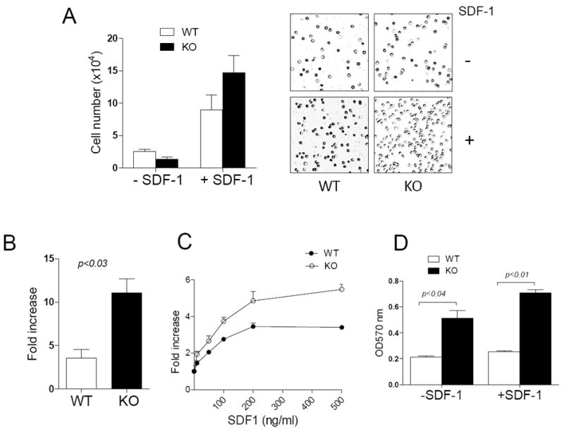
Figure 1. MIM-/- BM cells had a higher motility in response to SDF-1 than did WT cells.

(A) WT and MIM-/- (KO) BM cells were plated in the upper chamber of Transwell plates in the presence or absence of 200 ng/ml SDF-1. After 4h, cells migrated into the lower chamber were counted (left) or photographed using a 10 x objective lens (right). (B, C) The motility of cells toward either 200 ng/ml SDF-1 (B) or SDF-1 at different concentrations (C) was measured as above. The fold increase in the mobility was calculated by normalizing the number of mobilized cells to that measured in the absence of SDF-1. (D) BM cells were plated on 24-well plates pre-coated with 2 μg/ml fibronectin and incubated for 20 min. Attached cells were stained with crystal violet and quantified based on absorption at OD570nm. All the data represent mean ± SEM (n=3).
