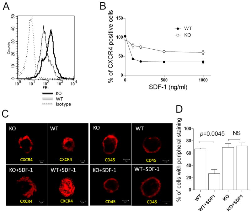
Figure 2. MIM-/- cells had increased in expression of CXCR4 on the surface.

(A) Freshly isolated BM cells were stained by PE-CXCR4 antibody and subsequently analyzed by flow cytometry. (B) BM cells were treated with SDF-1 for 10 min at concentrations as indicated, and then analyzed by flow cytometry for the surface expression of CXCR4. All the data represent mean ± SEM (n=3). (C) BM MNCs were stained with antibody against either CXCR4 or CD45 with or without SDF-1 treatment and examined by confocal microscopy. Bar, 2 μm. (D) Quantification of BM MNCs that showed CXCR4 staining mainly at peripheral areas.
