
Figure 3. MIM-/- BM cells increased in homing during BM transplant.
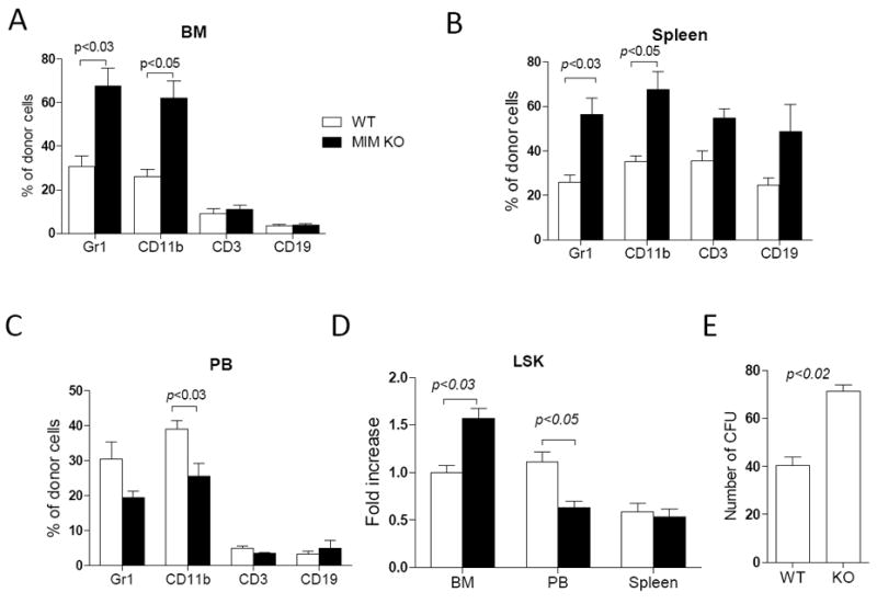
2×10⁶ WT or MIM-/- BM cells from CD4.2 mice were transplanted into lethally irradiated CD45.1 WT mice. The frequency of Gr1, CD11b, CD3 and CD19 cells in the donor population were determined by flow cytometry in the BM (A), the spleen (B) and the PB (C) of the recipients 24h after transplantation. Donor Lin-Scal-1+c-Kit+ (LSK) cells in the BM, the spleen and the PB of the recipients were also measured and normalized to WT donor LSK cells in the BM (D). All the data represent mean ± SEM (n=5). (E) BM cells were isolated from recipients 24 h after transplantation and analyzed by colony-forming assay (n=3). All p values were calculated by Student’s t-test.