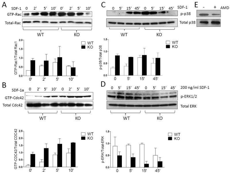
Figure 5. MIM-/- BM cells have enhanced CXCR4 signaling.

BM cells derived from WT and MIM-/- mice were treated with 200 ng/ml SDF-1 for the times as indicated and then analyzed for the presence of GTP-Rac (A), GTP-Cdc42 (B), phosphorylated p38 (C) and phosphorylated ERK1/2 (D) by Western blot. The charts below each image were the quantification results of three independent experiments. (E) Arresting BM cells of MIM-/- mice were treated with and without AMD3100 for 1h. The phosphorylated p38 was analyzed by Western blot.
