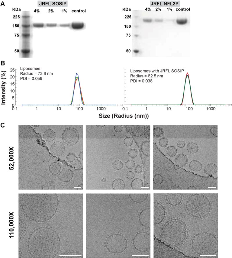
Figure 2. Characterization of JRFL SOSIP-conjugated liposomes.

(A) Reducing SDS PAGE of 4%, 2% and 1% Ni DGS-NTA(Ni) JRFL SOSIP and JRFL NFL trimer-conjugated liposomes. JRFL SOSIP and JRFL NFL2P soluble trimeric glycoproteins are included as controls. (B) Dynamic light scattering (DLS) of the 4% DGS-NTA(Ni) liposomes and JRFL SOSIP-conjugated liposomes was performed using a using Zetasizer Nano instrument to measure particle size and the polydispersity index. (C) Cryo-EM images of 4% Ni JRFL SOSIP liposomes at 52,000 and 110,000× magnification. Scale bar = 100 nm. See also Figure S1.
