
Figure 4. Binding of HIV-1 antibodies to JRFL SOSIP trimer-conjugated liposomes and soluble JRFL SOSIP trimer assessed by Bio-Layer Interferometry using Octet and negative stain EM.
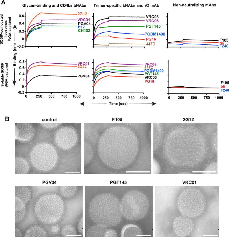
(A) JRFL SOSIP trimer conjugated to 4% DGS-NTA(Ni) liposomes (equivalent to 75 nmoles of phospholipids) or JRFL SOSIP trimers (10 mg/ml) were immobilized on WGA-captured streptavidin sensors and 20 mg/ml monoclonal antibodies (IgGs) were used as analyte. (B) 2% DGS-NTA(Ni) liposomes conjugated to JRFL SOSIP were incubated with 10 molar excess of respective IgG mAbs at 37°C for 30 min, stained with phospho-tungstate, viewed by EM and images were obtained with a CCD camera. All images are at 180,000× magnification. Scale bar = 100 nm. See also Figure S3.