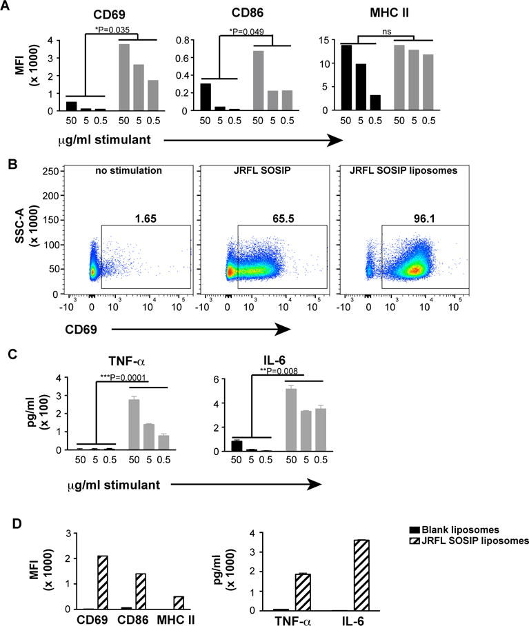
Figure 5. Activation of primary B cells by soluble JRFL SOSIP trimers and JRFL SOSIP trimer-conjugated liposomes.

B cells from b12 mature knock-in mice were negatively selected from splenocytes and induced by overnight incubation with either soluble JRFL SOSIP trimers or 4% liposomes conjugated with JRFL SOSIP trimers. The cell-surface activation markers and the cytokines secreted by the activated cells were analyzed by cell-surface staining or ELISA. (A) FACS staining of cell-surface activation markers plotted as MFI values. Soluble JRFL SOSIP (black); JRFL SOSIP conjugated to liposomes (grey bars). (B) Frequency of CD69+ cells upon activation by 50 μg/ml of soluble trimers or JRFL SOSIP trimer-conjugated liposomes. (C) TNF-α and IL-6 levels present in the supernatants of the B-cells upon overnight activation by soluble JRFL SOSIP trimers (black bars) or JRFL trimer-conjugated liposomes (grey bars) B cells were assessed. (D) MFI values of cell surface activation markers and levels of cytokines produced by B cells upon activation by 50 μg/ml of JRFL SOSIP liposomes or similar dilution of blank liposomes without any trimers on the surface. Statistical comparisons between groups are performed by paired t-test. See also Figure S4.
